

Chun Chen, Heng-Bang Wang, Ying-Ying Wu, Jian-Hua Xu*
Institute of Clinical Pharmacological, Fujian Medical University Fuzhou 350004, China
Ye Li, Zhi-Yong Xiao
Fujian Xianzhilou Biological Science & Technology Co., Ltd
ABSTRACT
OBJECTIVE To evaluate the inhibitory effects of Ganoderma extracts and spores oil on tumor cells in vitro and in vivo, and to investigate the possible mechanisms by observing their effects on topoisomerases and theeffect of Ganoderma spores’ oil on cell cycle.
METHODS Tumor cytotoxicity was detected by MTT method and Trypan blue staining method in vitro study and by mice xenografts as models’ in vivo study. Their effects on immunity indexes of mice bearing tumorwere also evaluated. The inhibitory effects on topoisomerases were measured by supercoiled DNA relaxationassay. Cell cycle was analyzed by flow cytometer. The level of cyclin D1 and cyclin E were observed by Western.
RESULTS The IC50 values of Ganoderma extracts on HL60, K562 and SGC-7901 for 24h were 0.44mg/ml, 0.39mg/ml and 0.90mg/ml; as for Ganoderma spores oil, they were 1.13mg/ml, 2.27mg/ml and 6.29mg/ml, respectively. In vivo study, the inhibitory rate of Ganoderma extracts (4g/kg/d, ig) on S180 and H22 were 39.1% and 44.6% of Ganoderma spores oil (1.2g/kg/d, ig) were 30.9% and 44.9%; As for Extract 1g/kg and2g/kg group, the inhibitory rate on S180 were 15.6% and 19.0%, on H22 were 28.8% and 38.5%. Ganoderma extracts had a certain effect in improving immune function of mice bearing tumors, while Ganoderma sporesoil had no significant effect on immunity indexes of mice. Both Ganoderma extracts and spores oil inhibitedactivities of topoisomerase I and II. Ganoderma spores oil was shown to be capable of blocking the cell cycle atthe transition from G1 to S phase and inducing a marked decrease of cyclin D1 level in K562 cells, with nosignificant change in cyclin E level.
CONCLUSION Ganoderma extracts and spores oil possess definite antitumor effects both in vitro and in vivo study. The antitumor mechanisms of both two are all related to their inhibitions on topoisomerase I and IIactivities, and for Ganoderma spores oil, the anti-tumor effects might also relate to the decrease of cyclin D1level which induced a G1 arrest of cell cycle.
KEY WORDS Ganoderma extracts; Ganoderma spores oil; tumor; topoisomerase I and II; cell cycle; cyclin D1
INTRODUCTION
Ganoderma lucidum (Ganoderma or Lingzhi) is one of most frequently used fungi in Chinese medicine. Modern pharmacological and clinical study had confirmed that Ganoderma contained abundant biological activity substances in its fruiting body, mycelia, and spore, and possessed variable functions such as immunomodulation, anti-aging, reducing blood lipid, anti-virus, and anti-tumor activities [1-6]. We had used multiple methods to extract activity ingredients from the fruiting body and spores of Ganoderma, including aqueous extract and ethanol extract of Ganoderma fruiting body, and Ganoderma spores oil which was extracted from broken spores by supercritical CO2 extraction technology. In this work, we examined the anti-tumor activity of mixture of aqueous extract and ethanol extract of Ganoderma fruiting body (hereinafter short for Ganoderma extracts) and Ganoderma spores oil, the influences on cell cycle and the effects on topoisomerases were also investigated.
METHODS
Preparations of Ganoderma extracts and spores oil
Ganoderma extracts and Ganoderma spores oil was provided by Fujian Xianzhi Lou Biological Science & Technology Co., Ltd (batch number: 20060901). Ganoderma extract, a brown powder, was dissolved in double distilled water to prepare different concentration solutions which were brown suspension. Ganoderma spores oil was a soft capsule with 0.5g/0.5ml golden oil in each capsule. The stock solution of Ganoderma spores oil was prepared by double distilled water which containing 6μl/ml(v:v) Tween 80.
Cell lines and culture
Human acute myeloid leukemia HL-60 cell line, human chronic myeloid leukemia K562 cell line, human gastric carcinoma cell line SGC 7901, murine S180 sarcoma cell line and murine H22 hepatoma cell line was all purchased from Shanghai Cell Institute of the Chinese Academy of Sciences. All tumor cells were maintained in RPIM 1640 (Gibco, USA) medium supplemented with 10% fetal calf serum and antibiotics (100kU/L penicillin and 100mg/L streptomycin) at 37°C in a humidified atmosphere of 5% CO2.
MTT assay
The cell cytotoxicity of Ganoderma spores oil in vitro was detected by MTT assay. MTT assay was based on the ability of live cells to utilize thiazolyl blue and convert it into dark blue formazan. Exponentially growing cells (2×10⁴/well) were seeded in 96 well plates and treated with a series of concentration of Ganoderma spores oil for 24 hours. Control cells were exposed to double distilled water containing the same concentration of Tween 80. And then 20μI of 5mg/mL MTT (Sigma, USA) were added to each well and incubates for 4 hours. The dark blue formazan crystals were solubilized in lysis buffer (10%SDS, 5% isobutyl alcohol, 12mmol/L HCl) and the optical density was detected at 492nm with a spectrophotometer. Growth inhibition rate was calculated according to the formula:
Inhibition rate = (1-mean OD value of treated group/mean OD value of control group) ×100%
IC50 value was calculated by linear regression method.
Trypan blue exclusion method
The cell cytotoxicity of Ganoderma extract in vitro was assessed by Trypan blue exclusion method. Exponentially growing cells were treated with a series of concentration of Extract of Ganoderma for 24 hours. Control cells were exposed to double distilled water. Then cells were dyed with 0.4% trypan blue. Viable cells (trypan blue dye-excluding) were counted using a light microscope. Inhibition rate was calculated according to the formula:
Inhibition rate = (1-viable cells in treated group/viable cells in control group) ×100%
Anti-tumor effects in vivo
Kunming mice (male and female, body weight 18~20g) were obtained from animal centre of Fujian Medical University. Tumor cells (S180 or H22) suspension (1×10⁶ cells in 200μl solution) was carefully inoculated intradermally into the left axilla of mice. Then, the tumor-bearing mice were randomly assigned to treatment experimental groups (ten mice in each group). Twenty-four hours after the tumor inoculation, mice were intragastric administrated with ①Extract of Ganoderma 4g/kg, ②Extract of Ganoderma 2g/kg, ③Extract of Ganoderma 1g/kg, ④Ganoderma spores oil 1.2g/kg, ⑤5-FU 25mg/kg (as a positive control), or ⑥0.9% NaCl solution 0.2ml/10g (as a negative control) once daily for 10 consecutive days. At the end of the experiments (day 12), mice were weighted and then sacrificed, the tumors, mice thymus gland and spleen were excised carefully and weighted.
Tumor suppression rate = (1-mean tumor weight in treated group/mean tumor weight in control group) ×100%
Thymus index = 100×thymus weight/body weight
Spleen index = 100×spleen weight/body weight
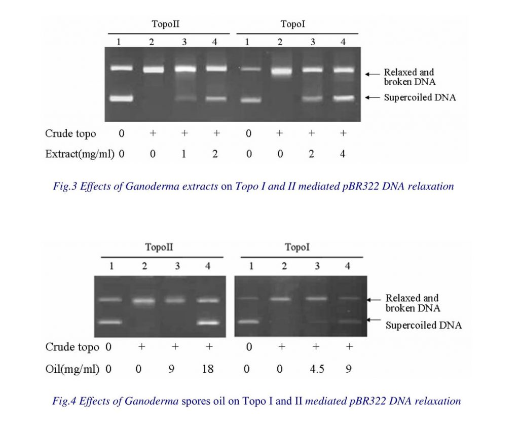
Catalytic Activity of Topoisomerases [7]
K562 cells (1 X 10⁷) were washed with PBS and then treated with lysis buffer (20 mM Tris, 1mM EGTA, 25 mMKCl, 5 mM MgCl2, 250 mM sucrose, and 0.5% NP40, pH 7.2) for 10 min at 4˚C, then centrifuged at 6000 rpm for 2min. The pellet was resuspended in 50μl of extraction buffer (20 mM Tris, 1 mM EGTA, 2 mM EDTA, 2 mM DTT, and 400 mM NaCl, pH 7.2) and incubated for 30min at 4˚C. After centrifuged at 14,000 rpm for 15 min, the supernatant (containing extracted topoisomerases) was obtained, and its protein concentration was determined by the procedure of Bradford before storage at 20°C.
The catalytic activity of topoisomerase II in extracts was evaluated as follows: the total reaction volume was 10μl, containing 2.5μl 4× reaction buffer (50 mM Tris pH 8.0, 120mM KCl, 0.5mM DTT, 0.5 mM ATP, 10 mM MgCl2, 30μg /ml BSA), 0.1mM ATP (Promega, USA), 0.2μg pBR322DNA (Promega, USA), different amount of topoisomerase II-containing crude extract, using double distilled water to make up10μl. After incubation at 37°C for 30 min, the reaction was stopped by the addition of 5μl of a solution maintained at 4°C containing 50 mM EDTA, 50% glycerol, and 0.25 mg/ml bromophenol blue. Samples were separated through a 1% agarose gel. After staining with ethidium bromide, gels were photographed under UV illumination and analyzed by SynGene GeneSnap Tools software.
The catalytic activity of topoisomerase I in extracts was evaluated as follows: the total reaction volume was 10μl, containing 1μl 10× reaction buffer (10mM Tris-HCl pH 7.9, 0.15 M NaCl, 1 mM EDTA, 0.1 mM spermidine, 0.1% BSA, 5% glycerol), 0.2μg pBR322DNA, different amount of topoisomerase I-containing crude extract, using double distilled water to make up10μl. The rest of procedures were the same as “topo II” method.
The inhibitory effects of Ganoderma extracts and Ganoderma spores oil on topoisomerase I and II was then detected. Selecting the amount of crude extract that exactly unwinding 0.2μg pBR322DNA to establish topoisomerase I and topoisomerase II reaction systems as mentioned above, then 2μl drugs was added in these two reaction systems and using double distilled water to make up10μl respectively. After incubation at 37°C for 30 min, the reaction was stopped and separated through agarose gel. The staining gel was analyzed under UV illumination.
Cell cycle analysis
The cell cycle was analyzed by flow cytometry. K562 cells were seeded in 6- well plates at a density of 1×10⁵/mL and treated with 1.25 and 2.5mg/ml Ganoderma spores oil for 12h. Then cells were washed twice with PBS and fixed in 70% ethanol at 4°C overnight. Prior to analysis, cells were washed and resuspended in PBS, then incubated with 10 mg/mL RNase A for 3~5 min and 50 mg/mL propidium iodide (Sigma, USA) at 4 °C for 30 min in a dark chamber. The cell cycle distribution (percentage of cells in different phases) was analyzed by flow cytometry (Epics XL, Coulter, USA) using Multicycle software.
Western blot analysis
K562 cells (1×10⁷) were washed with PBS and then treated with lysis buffer (50mmol/L Tris-HCl pH 8.0, 150mmol/L NaCl, 1mmol/L PMSF, 1mmol/ L Aprotinin, 1%NP-40) for 30 min at 4°C. After centrifuged at 10,000rpm for 20 min and protein concentration in the supernatant was determined. Equal amounts (30μg) of protein were separated by SDS-polyacrylamide gel electrophoresis and then electro- transferred onto nitrocellulose membranes in transfer buffer. The membrane was blocked with 5% BSA for 2h and then incubated with primary antibodies (cyclin D1, cyclin E and actin, purchased from Santa Cruz, USA) in blocking solution at 4°C for 2h, followed by extensive washing with PBS twice. Membrane was then incubated with alkaline phosphatase conjugated secondary antibodies in blocking solution at room temperature, followed by washing with TBS three times. The target proteins became visible following the addition of Alkaline Phosphatase Substrate Solution BCIP/NBT (Promega, USA).
Statistical analysis
Statistical analysis of the data was performed with t-test. Data were expressed as mean±SD. Differences with P<0.05 were considered statistically significant.
RESULTS
Inhibitory effects on tumor cells in vitro
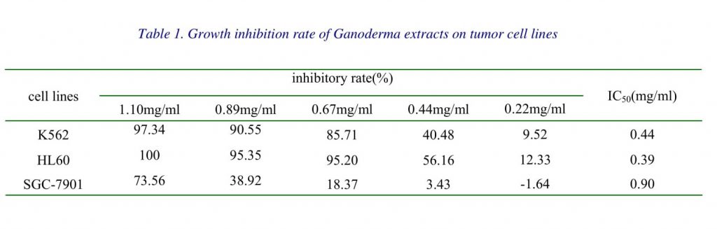
Anti-proliferative activities of Ganoderma extracts and Ganoderma spores oil on K562, HL60and SGC-7901 cell lines were determined. Usingtrypan blue exclusion method, dose-dependentinhibitions were observed when Ganoderma extractsconcentrations in the range of0.22~1.10mg/ml. IC50 value of Ganoderma extractson K562, HL60 and SGC-7901 cells were0.44mg/ml, 0.39mg/ml and 0.90mg/ml respectively (Table1).
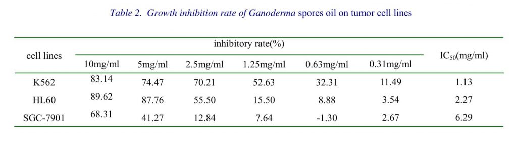
Using MTT assay, it was shown that treatment with Ganoderma spores oil concentrations of 0.31~10mg/ml caused dose dependent cytotoxicity in K562, HL60 and SGC-7901 with IC50 value of 1.13mg/ml, 2.27mg/ml and 6.29mg/ml respectively (Table 2)
Suppression of tumor growth in vivo
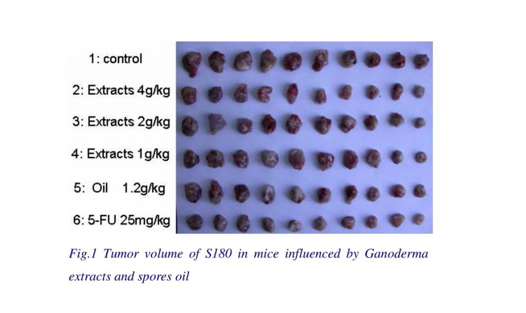
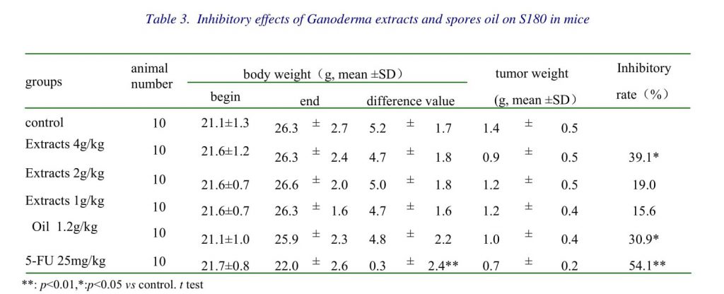
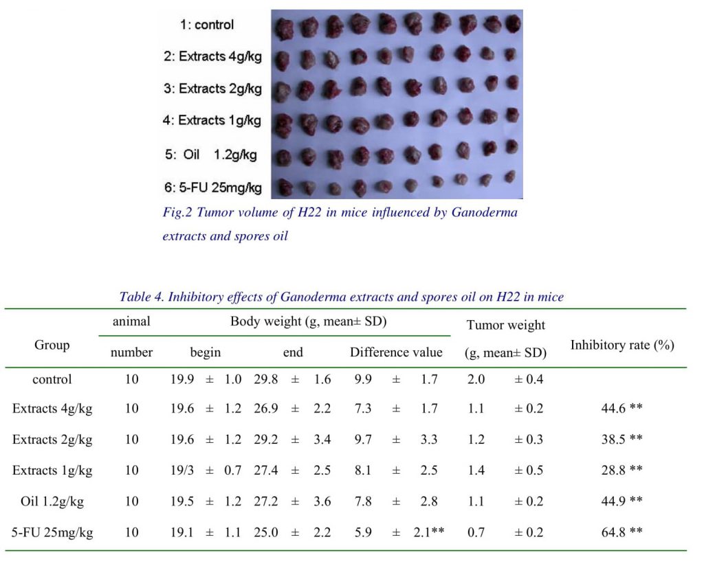
The effects of intragastric administration of Ganoderma extracts and Ganoderma spores oil on S180 in mice were examined (Fig.1 and Table3). Tab.3 showed that Extracts 4g/kg group and Oil 1.2g/kg group resulted in significant inhibitions of tumor growth by comparing the average tumor weight with control group at the end point of the experiment (P<0.05), while the inhibitory rates of Extracts 1g/kg and 2g/kg group were 15.6% and 19.0% (P>0.05). The average body weights were not significantly different among treatment groups only excepted 5-FU group. The suppression effects of Ganoderma extracts and Ganoderma spores oil on H22 in mice were shown in Fig.2 and Tab.4. Data in Tab.4 showed that both Ganoderma extracts and Ganoderma spores oil exhibited relatively strong inhibitory effects on H22 in mice. The inhibitory rate of Extracts 1g/kg, 2g/kg, and 4g/kg groups on H22 were 28.8%, 38.5% and 44.6%, respectively (P<0.01 compared with control group), and of Oil 1.2g/kg group was 44.9% (P<0.01). The average body weights were significantly decreased in Extracts 4g/kg group (P<0.05) and 5-FU group (P<0.01) when compared with control group.
Influences on immunity indexes of mice bearing tumors
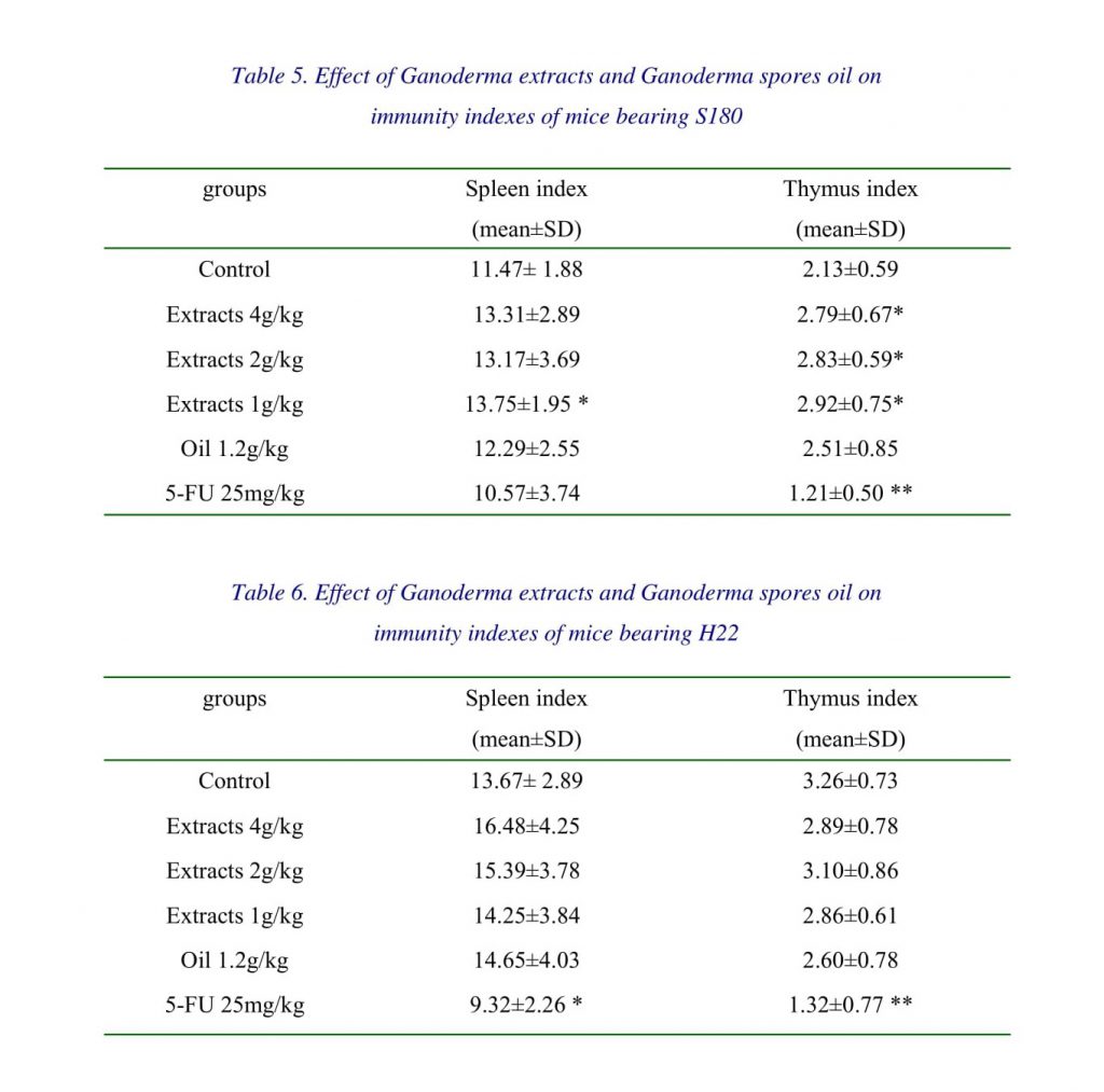
Table 5 showed that Ganoderma extracts increased the immunity indexes of mice bearing S180: the spleen index of Extracts 1g/kg group was increased significantly as compared with control group (p<0.05), the thymus indexes in Extracts 1g/kg, 2g/kg and 4g/kg groups were all improved significantly (p<0.05), while in 5-FU group was decreased significantly (p<0.01). Ganoderma spores oil exerted no significant effects on spleen index and thymus index.
Being treated with Extract of Ganoderma and Ganoderma spores oil, there was no significant change in the immunity indexes of mice bearing H22 (Table 6).
Inhibitory effects on topoisomerases activities
The activities of topoisomerases I and II are determined by the conversion of a fixed amount of supercoiled DNA into relaxed and broken DNA. Topoisomerase inhibitors could decrease the amount of relaxed and broken DNA and increased supercoiled DNA by inhibiting the catalytic activities of topoisomerases. The minimal concentration of crude topoisomerases from K562 required to fully relax 0.2μg pBR322DNA was used to test Ganoderma extracts and Ganoderma spores oil.
Our results showed that both of Ganoderma extracts and spores oil inhibited activities of topoisomerase I and II. Treated with Ganoderma extracts 2mg/ml, topoisomerase I and II had been all inhibited as reflected by the increased amount of supercoiled DNA and decreased amount of relaxed and broken DNA (Fig.3). When added Ganoderma spores oil 4.5 mg/ml into the reaction system of topoisomerase I, supercoiled DNA was observed. As the concentration of Ganoderma spores oil raised, a dose dependent manner was observed. By using Ganoderma spores oil 18mg/ml, the activity of topoisomerase II was then suppressed (Fig.4).
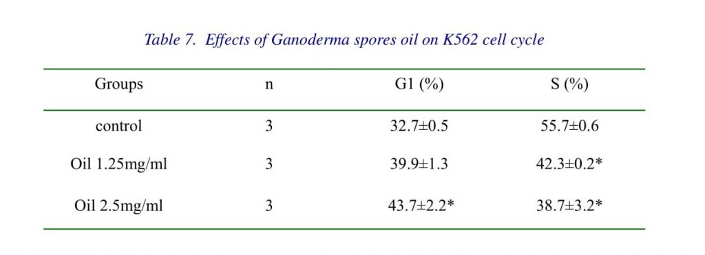
Influence of Ganoderma spores oil on cell cycle
The cell cycle was analyzed by flow cytometry. As shown in Table7, Ganoderma spores oil induced a G1 arrest in K562 cells. With the treatment of Ganoderma spores oil 1.25mg/ml for 12h, the percentage of S cells in K562 was decreased from 55.7±0.6 to 42.3±0.2 (p<0.05); when the concentration of Ganoderma spores oil was raised to 2.5mg/ml, the percentage of S cells was further decreased to 38.7±3.2 (p<0.05), with the percentage of G1 cells rose from 32.7±0.5 to 43.7±2.2 (p<0.05).
Results of Ganoderma spores oil on cyclin D1 and cyclin E
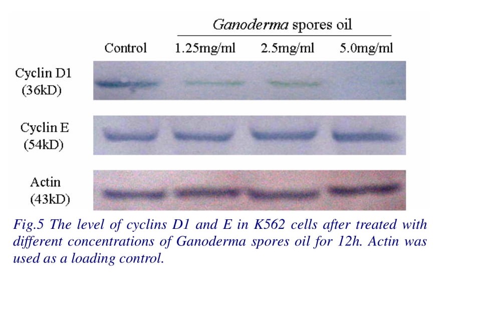
Cyclin D1 and E are important regulators of G1-S phase cell cycle transition. The expression levels of cyclin D1 and E were determined by Western blotting. The results showed that after the treatment of Ganoderma spores oil 1.25~5.0mg/ml for 12h, cyclin D1 expression in K562 decreased, but cyclin E expression had no obvious change (Fig.5).
DISCUSSION
Recently the effect of Ganoderma on tumors was regarded increasingly. Our work showed that both Ganoderma extracts and Ganoderma spores oil inhibited growth of human leukemia cells (K562 and HL60) and human gastric carcinoma cell (SGC-7901) in a dose-dependent manner, and significantly suppressed growths of transplant tumor of S180 and H22 in mice. Ganoderma extracts and Ganoderma spores oil possess definite antitumor effects both in vitro and in vivo study.
Since ancient time, Ganoderma has been widely used as a popular herbal medicine for the promotion of health. Until now there were many reports concerned with the immunomodulatory activities of Ganoderma [8-9]. By detecting the immunity indexes of mice bearing S180 or H22, we concluded that Ganoderma extracts had a certain effect in improving immune function, while Ganoderma spores oil had no significant effect on spleen index and thymus index of mice. One of main components of Ganoderma extracts is polysaccharides which has been reported as immune function enhancer [8-11]. As there were little polysaccharides (water-solubility substance) in Ganoderma spores oil, it exhibited no obvious effect on immunity. Our work also indicated that the anti-tumor effects of Ganoderma were safer than that of 5-FU which decreased body weight and immunity indexes of mice (Table1 and Table 2).
To investigate the possible mechanism of Extract of Ganoderma and Ganoderma spores oil, we observed their effects on topoisomerases and the effect of Ganoderma spores oil on cell cycle.
DNA topoisomerases are a class of enzymes involved in the regulation of DNA supercoiling. Type I topoisomerases change the degree of supercoiling of DNA by causing single-strand breaks and religation, whereas type II topoisomerases cause double-strand breaks. Both activities are especially crucial during DNA transcription and replication, when the DNA helix must be unwound to allow proper function of large enzymatic machinery. Topoisomerases overexpression have been linked to several human malignancies and are the target for many chemotherapeutic agents [12]. If topoisomerases are blocked, the cell will encounter problems during transcription of the DNA and during cell division. Cancer chemotherapy takes advantage of this, using drugs that block topoisomerases to kill rapidly dividing cancer cells. For instance, the widely used anthracycline drugs, like doxorubicin and daunorubicin, attack class II topoisomerases, and the plant toxin camptothecin blocks the relaxing action of class I topoisomerases.
Ding J [13] et al reported that China Ganoderma lucid essence (containing Ganoderma Lucidum extract powder and sporoderm-broken spores powder) could inhibit the activities of topoisomerase I and II. Consistence with these reports, our results showed that the catalytic activities of topoisomerases I and II were all inhibited by both Ganoderma extracts and Ganoderma spores oil. This suggested that topoisomerase I and II might be target of the actions of Ganoderma extracts and spores oil.
Extracting Ganoderma spores oil from broken spores by supercritical CO2 extraction technology is developed in recent year, and several recent studies had concerned about the anti-tumor effect of Ganoderma spores oil [14-15]. In our work, a G1 arrest was detected in K562 cells treated with Ganoderma spores oil (Ganoderma extracts was a brown suspension and can’t detect by flow cytometry). Ganoderma spores oil mainly contains triterpenes, fatty acids, et al. Tang W [16] et al reported that Ganoderic acid T (a triterpenoid chemical) markedly inhibited the proliferation of a highly metastatic lung cancer cell line by apoptosis induction and cell cycle arrest at G1 phase. So, we presumed that the cell cycle blocking effect of Ganoderma spores oil was mainly related with triterpenes.
To our knowledge, this paper described for first time that the level of cyclin D1 decreased in Ganoderma spores oil treated K562 cells. Cyclin D1 plays a critical role in the G1~S transition of cell cycle and induces a G1 arrest when its level is too low. It might therefore be suggested that the decrease of cyclin D1 is one of the reasons for the G1 arrest of the K562 cell cycle, which resulted in the inhibition of cell growth in the presence of Ganoderma spores oil.
The active substances in Ganoderma are worth further studying.
CONCLUSIONS
Ganoderma extracts and spores oil possess definite antitumor effects both in vitro and in vivo study. The antitumor mechanisms of both two are all related to their inhibitions on topoisomerase I and II activities, and for Ganoderma spores oil, the anti-tumor effects might also relate to the decrease of cyclin D1 level which induced a G1 arrest of cell cycle.
REFERENCES
1. Mizuno T, Wang G, Zhang J, et al. Reishi, Ganoderma lucidum and Ganoderma tsugae: bioactive substances and medicinal effects. Food Rev. Int. 1995, 11:151-66.
2. WD Li, Q Han, ZB Lin. Study on Effect of Ganoderma Polysaccharide and Lentinan in Adjusting Cyclophosphamide Induced Immunosuppression in Tumor Bearing Mice. Chinese Journal of Integrated Traditional and Western Medicine, 2001, S1:50.
3. Hong KJ, Dunn DM, Shen CL, Pence BC. Effects of Ganoderma lucidum on apoptotic and anti-inflammatory function in HT-29 human colonic carcinoma cells. Phytother. Res. 2004, 18:768-70.
4. Komoda Y, Shimizu M, Sonoda Y, Sato Y. Ganoderic acid and its derivatives as cholesterol synthesis inhibitors. Chem. Pharm. Bull. 1989, 37:531-3.
5.Min BS, Nakamura N, Hattori M. Triterpenes from the spores of Ganoderma lucidum and their inhibitory activity against HIV-1 protease. Chem. Pharm. Bull. 1998, 46:1607-12.
6. Min BS, Gao JJ, Nakamura N, Hattori M. Triterpenes from the spores of Ganoderma lucidum and their cytotoxicity against meth-A and LLC tumor cells. Chem. Pharm. Bull. 2000, 48:1026-33.
7. Anna Kruczynski, Jean-Marc Barret, Benoit van Hille, et al. Decreased nucleotide excision repair activity and alterations of topoisomerase IIα are associated with the in vivo resistance of a P388 leukemia subline to F11782, a novel catalytic inhibitor of topoisomerases I and II. Clinical Cancer Research. 2004, 10:3156-68.
8. LS Lei, ZB Lin. Effect of Ganoderma polysaccharides on T cell subpopulations and production of interleukin 2 in mixed lymphocyte response. Acta Pharmaceutica Sinica. 1992, 05:2.
9. YH Hu; ZB Lin; YQ He, et al. Polysaccharides isolated from mycelia of Ganoderma lucidum induced on HL 60 cell apoptosis by enhancing macrophage activity. Chinese pharmacological bulletin. 1999, 1:8.
10. XL Zhu, ZB Lin. Effects of Ganoderma lucidum polysaccharides on proliferation and cytotoxicity of cytokine induced killer cells. Acta Pharmacologica Sinica. 2005, 26 (9):1130-37.
11. Gao Y, Gao H, Chan E, et al. Antitumor activity and underlying mechanisms of ganopoly, the refined polysaccharides extracted from Ganoderma lucidum in mice. Immunological Investi- gations. 2005, 34(2):171-98.
12. Nathalie Dias, Hervé Vezin, Amélie Lansiaux and Christian Bailly. Topoisomerase Inhibitors of Marine Origin and Their Potential Use as Anticancer Agents. Topics in Current Chemistry[S], Springer Berlin / Heidelberg, 2005, Volume 253: 89-108.
13. Chao J, Chen Q, Ding J, et al. Inhibitory effects of China Ganoderma lucid essence (CGLE) on DNA topoisomerases and its ability to induce apoptosis of K562 cell. Chinese Journal of Cacer.1999, 18:661-3.
14. Gao JJ, Min BS, Ahn EM, et al. new triterpene aldehydes, lucialdehydes A-C, from Ganoderma lucidum and their cytotoxicity against murine and human tumor cells. Chemical & Pharmaceutical Bulletin. 2002, 50(6):837-40.
15. Yuan YS. Anti-implanted hepatoma effects of Ganoderma spores oil. Anhui Medical and Pharmaceutical Journal.2005, 9 (3):173-4.
16. Tang W, Liu HW, Zhao WM, et al. Ganoderic acid T from Ganoderma lucidum mycelia induces mitochondria mediated apoptosis in lung cancer cells. Life Sciences. 2006, 80(3):205-11.
* Correspondence to Prof Jian-Hua XU
Phone: 86-591-83569307. Fax 86-591-22862016
Email: xjh@mail.fjmu.edu.cn