
Zhenya Zhang 1, Yan-ting Wang 2, Jiayi Cheng 2, Wei Zhang 3 *, Zhikun Wang 4
1The Fourth Hospital of Hebei Medical University, Shijiazhuang 050000, China
2Department of Chinese Materia Medica, Hebei Medical University, Shijiazhuang 050091, China
3Department of pharmacology, the school of basic Science, Hebei Medical University, Shijiazhuang 050091, China
4Department of Digestion, Hebei Traditional Chinese Medical hospital, Shijiazhuang 050011, China
ABSTRACT
OBJECTIVE To investigate the effect of supercritical fluid extract of Xiaoguishao on proliferation and apoptosis in human gastric cancer cell line BGC-832.
METHODS BGC-823 cells were treated with the supercritical extract of Xiaoguishao for 48h. Western Blotting was used to detect the protein expression of Bcl-2 and PCNA in cultured human gastric cell line BGC-832; Annexin V/PI double staining was used to detect cell apoptosis rate.
RESULTS Supercritical fluid extract of Xiaoguishao significantly increase the apoptosis rate of human gastric cancer cell line BGC-823 in vitro after 48 hours and inhibit the expression of Bcl-2 and PCNA.
CONCLUSION Supercritical fluid extract of Xiaoguishao can significantly inhibit the proliferation of human gastric cancer cell line BGC-823 and induce its subsequent apoptosis.
KEYWORDS Precancerous lesions of gastric cancer (PLGC); supercritical extract of Xiaoguishao; BGC-823; Bcl-2; PCNA; proliferation; apoptosis
INTRODUCTION
Precancerous lesions of gastric cancer (PLGC) refer to moderate and severe dysplasia and/or incomplete colon metaplasia. It has an increasing risk to develop into gastric cancer [1, 2]. Studies have shown that Bcl-2 oncogene is closely correlated with cell apoptosis. Its abnormal expression is found during the development of precancerous lesions of gastric cancer (PLGC) [2]. Overexpression of proliferating cell nuclear antigen (PCNA) is often found in the nuclei of proliferating cells, the detection of its expression in cell lines can be used as a marker for the evaluation of cellular proliferation. Clinical studies have shown that Xiaoguishao can be used to significantly improve clinical symptoms of PLGC, in addition, supercritical fluid extract of Xiaoguishao can significantly improve the PLGC symptoms in rats, which may be due to induction of antioxidant activities and other effects [3]. It also significantly inhibited the proliferation of human gastric cancer cell line BGC-823 in rats. Xiaoguishao mixture (XGS) is made from Chinese Angelica and Peony Powder (Danggui Shaoyao San and Xiaoxianxiong Tang). It has been used for more than twenty years in clinical practice for relieving symptoms of precancerous chronic atrophic gastritis, it was found to successfully improve the clinical symptoms. In the earlier stages of the current study, it was found that XGS can be broke down to the four Chinese herbs: Danggui (Radix Angelicae Sinensis; Chinese Angelica), Baishao (Radix Paeoniae Alba; White Peony Root), Baizhu (Rhizoma Atractylodis Macrocephalae; Largehead (Atractylodes Rhizome), Gualou (Fructus et Semen Trichosanthis Kirilowii; Mongolian Snake gourd Fruit) (1:1:1:2). Effective compounds from these herbs can be extracted using the supercritical fluid method. Based on previous clinical, in vivo, and in vitro studies, this paper aims to further investigate the effect of supercritical fluid extract of XGS on human gastric cancer line BGC-823 and Bcl-2 oncogene, PCNA, and cell apoptosis, furthermore, to understand mechanisms of the effect of XGS supercritical fluid extract on PLGC.
MATERIALS AND METHODS
Material
Human gastric cancer cell line BGC-823 obtained from Chinese Academy of Science Shanghai Institute of Cell biology. Supercritical CO2 extraction: (HA 121-50-02 Supercritical Extraction Equipment, Jiangsu Province Nantong Hua An Supercritical Equipment Co., Ltd.). In vitro culture, cell culture was divided once every 2-3 days; RPMI 1640 culture medium obtained from Gibco; fetal bovine serum obtained from Hangzhou Every Green Organism Engineering Materials Co., Ltd; BCA protein assay kit obtained from Pierce; mouse antihuman HRP IgG monoclonal antibodies for Bcl-2 and PCNA obtained from Beijing Zhongshan Biotechnology Co., Ltd; Tris obtained from Sigma; SDS obtained from Bion Biotechnology C., Ltd; Epies-XL II Flow Cytometer obtained from Beekman Coulter; DYY-III7B electrophoresis system and DYC-722A electrophoresis tank obtained from Beijing Liuyi Instrument Factory.
Methods
Extraction
Freeze-dried the herbs Danggui (Radix Angelicae Sinensis; Chinese Angelica), Baishao (Radix Paeoniae Alba; White Peony Root), Baizhu (Rhizoma Atractylodis Macrocephalae; Largehead Atractylodes Rhizome), and Gualou (Fructus et Semen Trichosanthis Kirilowii; Mongolian Snake gourd Fruit) using lyophilizer, then crushed to 75 particle size. The crushed particles were mixed to the ratio 1:1:1:2, and then placed into the extraction apparatus. Extraction condition: Extraction apparatus pressure at 25 Mpa, temperature at 40˚C; Isolation apparatus I pressure at 8Mpa, temperature 50˚C, Isolation apparatus II pressure at 6Mpa, temperature 50˚C; CO2 flow rate at 15L/h; extraction time 2 hours. Fluid extract of green color was dissolved in acetone and diluted with culture medium to make final concentrations of 12.5, 25, 50, 100, 200mg/kg of XGS extract respectively.
Cell Culture
Cell line BGC-823 was grown on RPMI 1670 culture medium containing 10% FBS at 37˚C and 5% CO2, cells grown until reaching logarithmic phase, then used 0.5% trypsin digestion and Trypsin Blue Staining to verify cell viability ≥95% and count cells for use.
Western blotting detection for protein expression of Bcl-2 and PCNA
Plated logarithmic phase BGC-823 cell line at 2X10⁵ cells per well in 6-well plates and grown for 4 hours, after which XGS extracts of final concentrations 0. 50, 100, 200 mg/kg were added. 48 hours after XGS treatment, cells were collected, centrifuged, and the supernatant discarded4. All groups of cells were then precipitated and extracted for protein. The protein amount was quantified, after which protein samples were ran on SDS-PAGE (sodium dodecyl sulfate polyacrylamide gel electrophoresis). Protein samples were loaded at 25μl per lane. Protein samples in the gel were then transferred to nitrocellulose membrane using electrophoretic transfer (Western blotting). The membrane was blocked using TBST buffer containing 1% Solcoseryl Protein at room temperature for 1.5 hours, and then left incubating in primary antibodies (dilution of 1:5000) at 4˚C overnight. After washing with TBST buffer three times, incubated again with secondary antibodies (IgG antibodies, dilution of 1:3000) at room temperature for 1 hour. After washing with TBST buffer three times again, the membrane was reacted with ECL (enhanced chemiluminescent) reagent for 1 minute. Positive bands were measured for optical density (OD) value. β-actin was used as the internal standard. The same β-actin immunoblot positive band OD detection procedure was used to calculate the ratio of Bcl-2, PCNA band OD value to β- actin band OD value, in order to quantify the relative expression of protein in each group.
Cell Apoptosis Detection
Plate logarithmic phase BGC-823 cell line at 2X10⁵ cells per well in 6-well plate at 37˚C and 5% CO2 for 4 hours, after which XGS extracts of final concentrations 0, 12.5, 25, 50, 100, 200 mg/L were added, treatment of each concentration was set for 4 replications (4 wells), 48 hours after treatment, cells were collected after trypsin digestion, and washed with PBS twice. Annexin V/PI double staining and FCM (flow cytometry) were conducted to measure cell death rate.
RESULTS
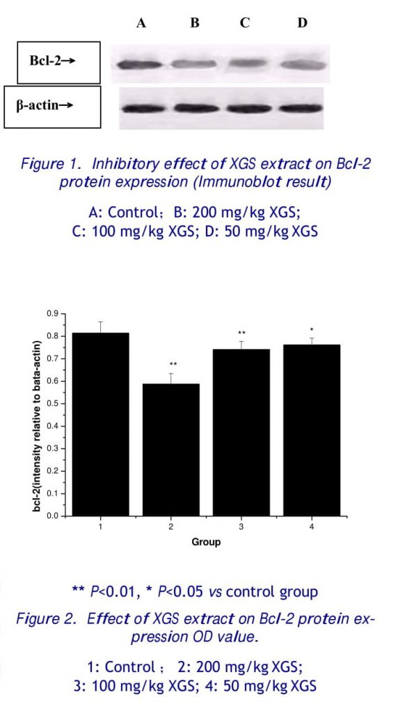
2.1 Effect of Xiaoguishao on Bcl-2 protein expression
The results show that three different concentrations of XGS extracts can significantly reduce protein expression of Bcl-2 in human gastric cancer cell line (BGC-823) 48 hours after treatment. There is statistically significant difference when compared to the control group (P<0.01 or P<0.05). Western blotting result is shown in Fig. 1, band OD values is shown in Fig 2.
2.2 The effect of supercritical extract of Xiaoguishao on the expression of the PCNA
The results show that XGS extracts at concentrations of 100 mg/kg and 200 mg/kg can significantly reduce protein expression in human gastric cancer cell line (BGC-823) 48 hours after treatment. There is statistically significant difference when compared to the control group (P<0.05). Western blotting result is shown in Fig. 3, band OD values is shown in Fig 4.
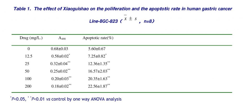
2.3 Cell Apoptosis
Drug treatment at different concentrations shows different proliferation and apoptotic rate (Table 1) in human gastric cancer line (BGC-823) 48 hours after treatment, the difference is significant when compared to the control solution group.
DISCUSSION
Impaired apoptosis may enhance tumor progression and promote metastasis. Bcl-2 (B Cell lymphoma/Leukemia-2) is one of the most important oncogenes found to inhibit tumor cell apoptosis. Bcl-2 promote development of carcinoma not by enhancing cell proliferation, but by inhibiting cell apoptosis and prolonging cell life span and in doing so cause accumulation of abnormal cells [5]. Bcl-2 is an oncogene found at the t (14,18) chromosomal translocation point and regulates apoptotic event by prolonging cell survival. Studies have shown Bcl-2 positivity to be 65.4% in human gastric cancer tissues, 44.2% in precancerous tissue, and almost none in normal gastric mucosal tissue, which seems to suggest expression of Bcl-2 to be closely related to the development of precancerous gastric lesions to gastric carcinoma [6, 7].
Proliferating Cell Nuclear Antigen (PCNA) acts as an auxiliary factor for DNA polymerase δ in eukaryotic cells. Its molecular weight is 36KD. PCNA is an antigen that is expressed in the nuclei of cells during the DNA synthesis phase of the cell cycle and reaches maximum level during S phrase. The changes in its quantification are the same as in DNA, measuring its cellular expression can be used as a marker for the evaluation of cellular proliferation. It is also one of the most useful markers for cell proliferation viability. Hence, PCNA can be used to evaluate proliferation rate of tumor cells, and the investigation of the proliferation state of tumor cells. Clinical studies have shown PCNA expression is gradually elevated in superficial gastritis, chronic atrophic gastritis, intestinal metaplasia, dysplasia, and gastric carcinoma [8].
Apoptosis is a form of programmed cell death that determines the cell morphology and survival. It is currently believed that induction of tumor cell apoptosis is the key to successful tumor therapy. Our studies have shown that XGS extracts at different concentrations can significantly inhibit the proliferation of human gastric cancer cell line and can induct apoptosis.
This study uses the supercritical CO2 extraction method. This method has strong solubility toward organic materials, in addition to good selectivity, and can complete the entire extraction-isolation procedure at near room temperature. It is particularly suitable for isolation of heat sensitive and chemically unstable nature products. Compared to traditional organic fluid extraction method, it is low-cost, easy to operate, and safe for extraction, in addition to giving high yield and few impurities, among other merits.
CONCLUSION
This study result shows XGS extracts can significantly inhibit the gene expression of Bcl-2 and PCNA, and furthermore from in vitro studies showing that XGS extracts can significantly induce apoptosis of tumor cells and inhibit progression of tumor carcinoma. Further studies on the extraction of effective components from XGS extracts and selection of drug activities are still under work.
REFERENCE
1. Ling H, Shao XL, Jian HW. Effect of “Wei Yan Xiao” on bcl-2 Oncogene mRNA Expression in Precancerous Lesion of Gastric Cancer, Shang Hai ZhongYi Yao Za Zhi 2006, 40(1):27-28
2. Hu L, Lao SX, Tang CZ. Expression of bcl-2 oncogene in gastric precancerous lesions and its correlation with syndromes in Traditional Chinese Medicine. Word J Gastroenterol 2005, 11(21): 3293-3296
3. Correa P, Miller MJ. Carcinogenesis, apoptosis, and cell proliferation. Br Med Bull 1998; 54: 151-162
4. Xu A, Li S, Liu J. Correlation between apoptosis and proliferation in gastric pre-carcinoma. Zhonghua Yixue Zazhi 1999; 79: 185-186
5. Wang ZK, Liu H, Zhang W. Clinical effect of granula of Xiaoguishao granula of traditional Chinese herbs on patients with precancerous lesions of gastric cancer. Hebei Journal of Traditional Chinese Medicine 2007, 29(6): 495-497
6. Lee PC, Salyapongse AN, Bragdon GA, Shears LL 2nd, Watkins SC, Edington HD, Billiar TR. Impaired wound healing, and angiogenesis in eNOS-deficient mice. Am. J. Physiol 1999, 277: H1600–1608
7. Liu HF, Liu WW, Fang DC. Expression of bcl-2 gene in gastric carcinoma and precancerous lesions and its regulatory role in apoptosis. Acta Academiae Medicinae Militaris Tertiae 2004, 26(7): 580-583
8. Ding SY, Yan LP, Shi F. Expression and significance of Bcl- 2 and Ki-67 protein in gastric carcinoma and its Para tissue Medical Journal of QiLu 2005, 20(2):126-129
9. Yoo J, Park SY, Robinson RA. Ras gene mutations and expression of Ras signal transduction mediators in gastric adenocarcinomas1 Arch Pathol Lab Med 2002, 126 (9):10962- 11001
Correspondence to: Wei Zhang
Foundation Item: This study was supported by the State Administration of Traditional Chinese Medicine of Hebei Province (No. 2007040)
Telephone: 0311-86265319; Email: zhangwei666cn@Yahoo.com.cn